Exosome Therapy for the Under-Eye Area
November 17, 2026
Can Spider Veins Return? Causes and Ways to Reduce Risk
November 19, 2026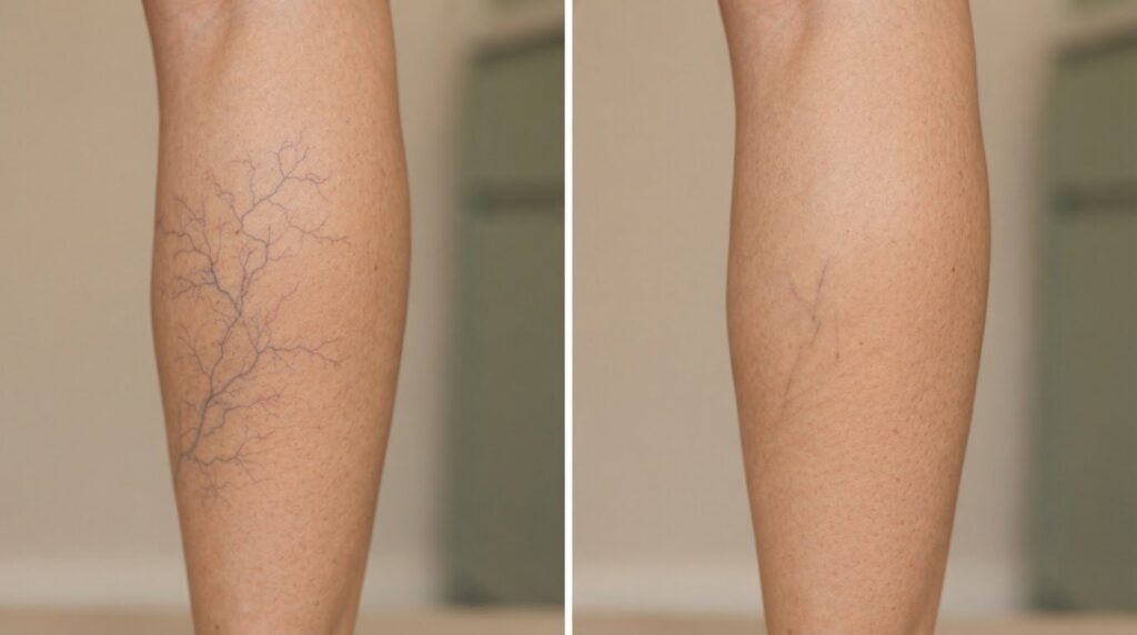
A before-and-after image is a snapshot, not a timetable. Sclerotherapy results before and after treatment can show spider veins beginning to improve within three to six weeks, while larger veins may take three to four months to respond. Treated veins may darken as the body gradually absorbs them, and some may remain visible.
It’s understandable to want a clear picture of what treatment can achieve. Results depend on the veins treated, whether underlying feeder veins are involved, and how healing progresses. This article explains what foam sclerotherapy examples can show, why results develop gradually, and how aftercare, compression stockings and further treatment may affect the outcome. Photographs taken at different follow-up points can capture very different stages of the same process.
Key Takeaways
- Compare before-and-after photos at similar angles and follow-up stages to better understand how vein changes develop.
- Follow the aftercare plan, including any compression instructions, as advised for your treatment.
- Consider how feeder veins beneath the skin may influence visible surface veins and the treatment approach.
- Foam sclerotherapy examples can help distinguish changes in appearance from changes in symptoms and show why treatment may be staged.
Before-and-after examples of varicose vein treatment
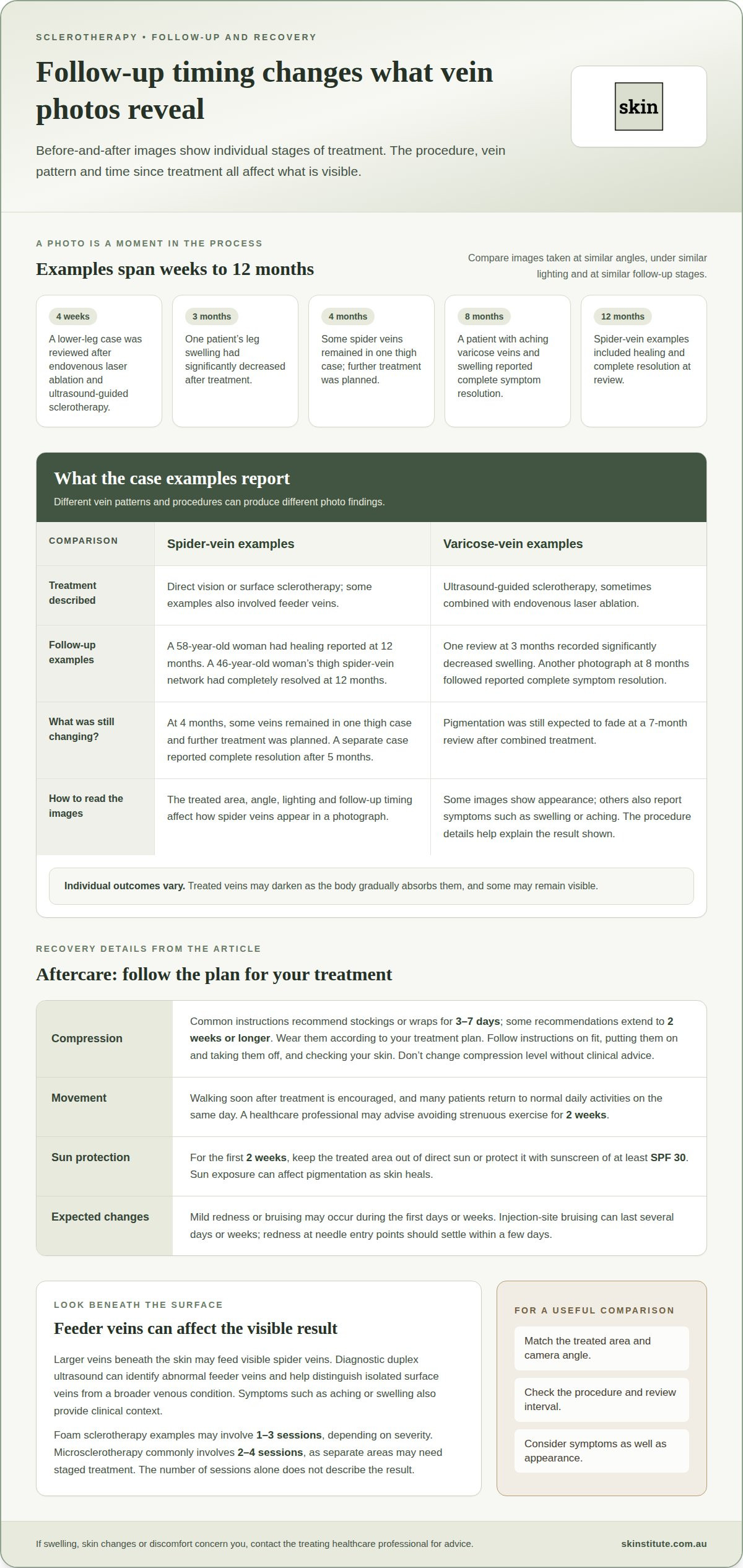
Before-and-after photos capture one point in a gradual treatment process. A 58-year-old woman with thigh spider veins had healing reported 12 months after treatment. In another case, a 46-year-old woman’s thigh spider-vein network had completely resolved 12 months after direct vision sclerotherapy. The examples show different individual outcomes at a similar follow-up stage.
Some varicose vein examples record symptoms as well as appearance. One patient with varicose veins, swelling and venous staining was reviewed three months after endovenous laser ablation and ultrasound-guided sclerotherapy. Leg swelling had significantly decreased. Another patient with aching varicose veins and swelling reported complete symptom resolution in a photograph taken eight months after treatment.
Spider vein treatment photos can show that results are still developing. One thigh case was photographed four months after bilateral ultrasound-guided and direct vision sclerotherapy. Some spider veins remained, and further treatment was planned. Photos are most useful to compare when the treated area, angle, lighting and follow-up timing are similar.
Some cases combine sclerotherapy with laser treatment. A lower-leg varicose vein case was reviewed four weeks after endovenous laser ablation and ultrasound-guided sclerotherapy. In a separate left-leg case, pigmentation was still expected to fade at a seven-month review after the same combined approach.
Laser treatment and sclerotherapy are distinct procedures. Some examples show sclerotherapy alone, while others include endovenous laser ablation. Knowing the procedure details helps explain what the photographs show. Sclerotherapy involves injecting a liquid into selected veins; laser treatment is a separate procedure that may be combined with it.
Medicare or private health insurance coverage in Australia depends on the circumstances and applicable benefits. Genuine Australian before-and-after photos can be found in clinical case examples that identify the treatment and follow-up timing. The examples described here include photographs at four weeks, four months and 12 months, illustrating why review timing matters.
Aftercare and compression stockings
After sclerotherapy, compression stockings or wraps support the treated area. Common instructions recommend wearing them for three to seven days. Other recommendations extend compression to two weeks or longer. The garment and wearing schedule should follow the treatment plan, not a general timeline. Compression can help manage local pressure and swelling as treated veins begin to close.
Stocking fit matters during daily use. A garment that slips, bunches or creates painful pressure can be difficult to wear correctly. Follow the clinician’s instructions for putting stockings on and taking them off, and for checking your skin. If the plan specifies continuous wear for an initial period, follow that schedule. Don’t change the compression level without clinical advice.
Movement is also part of routine recovery. Walking soon after treatment is encouraged, and many patients return to normal daily activities on the same day. A healthcare professional may advise avoiding strenuous exercise for two weeks. This allows ordinary movement while limiting strenuous activity during early healing. After an ultrasound-guided or endovenous procedure, follow the specific instructions for that session.
For the first two weeks, keep the treated area out of direct sun or protect it with sunscreen of at least SPF 30. Sun exposure can affect pigmentation as the skin heals. Mild redness or bruising may occur during the first days or weeks; injection-site bruising can last several days or weeks. Redness at needle entry points should settle within a few days.
Aftercare doesn’t make every visible change immediate. In one patient example, a photograph taken eight weeks after treatment showed a later stage than the initial recovery period. If swelling, skin changes or discomfort concern you, contact the treating healthcare professional for advice. Wear compression as instructed, and don’t treat it as a replacement for follow-up or review of the venous condition.
Feeder veins and underlying varicose veins
Visible spider veins can have larger feeder veins beneath the skin. These underlying veins may affect treatment planning and visible results. Diagnostic duplex ultrasound can identify abnormal veins feeding surface vessels. This assessment helps distinguish isolated spider veins from a broader venous condition. Symptoms such as aching or swelling also provide relevant clinical context.
In one case, a 56-year-old woman had visible spider veins and feeder veins. After surface sclerotherapy, both were reported as resolved at 12 months. Another 40-year-old woman had prominent feeder and spider veins. One direct vision sclerotherapy treatment was followed by complete resolution after five months. These examples show why follow-up timing matters.
Foam sclerotherapy is used in examples involving varicose veins. The foam is delivered into the vein, with ultrasound guidance where appropriate. Treatment may involve one to three sessions, depending on severity. The number of sessions alone doesn’t describe the result. Vein size, symptoms and the underlying pattern also matter.
Microsclerotherapy targets small surface spider veins, including fine red or blue vessels. Reticular veins are larger blue veins that can lie beneath spider veins. Their presence may change how a clinician treats the surface network. Microsclerotherapy commonly involves two to four sessions, as separate areas may need staged treatment.
Leg spider veins and blue veins can therefore reflect different vessel sizes and depths. A surface-only approach may not address an abnormal feeder vein. One 66-year-old woman reported worsening spider veins after surface sclerotherapy alone elsewhere. This example underlines the value of assessing the venous pattern before treatment. Compression may form part of the aftercare plan, but it doesn’t identify or treat feeder veins.
Examples of results after foam sclerotherapy
Foam sclerotherapy examples often include another procedure, such as endovenous laser ablation. The treatment details matter when interpreting each photograph. An image records appearance at one review point; it doesn’t show how much change occurred at other stages. Treated veins may darken within two weeks, while some remain visible after treatment.
One male patient’s varicose vein result was shown six weeks after endovenous laser ablation and foam sclerotherapy. Another male patient’s result was photographed five weeks after the same combination. A separate male patient had radiofrequency ablation with foam sclerotherapy, with a result shown six weeks later. These examples record follow-up timing, not a typical outcome for every patient.
Female patient examples also include both combined approaches. One result was shown eight weeks after endovenous laser ablation and foam sclerotherapy. Other reviews followed that combination at nine weeks. After radiofrequency ablation with foam sclerotherapy, one result was shown at six weeks and another at seven weeks. To compare these cases, note both the procedure and review interval.
The number of sessions for visible results varies by vein type and extent. Ultrasound-guided foam sclerotherapy for varicose veins may involve one to three sessions. Spider vein microsclerotherapy commonly involves two to four sessions. A review after one session may show remaining veins, so read photographs alongside the treatment plan.
Redness or small vessels may appear days or weeks after treatment. These changes can fade over the following months, while pigmentation may take longer. For a useful comparison, record the area, procedure and photo timing, then consider any remaining veins and skin changes. This gives a clearer picture than judging one image alone.
For a clinical assessment of visible veins and suitable treatment options, discuss sclerotherapy treatment.
Skinstitute: Sclerotherapy
Skinstitute provides sclerotherapy for people in Melbourne seeking treatment for visible spider or varicose veins. A face-to-face consultation helps assess the concern and inform a personalised treatment plan. Vein size, location, appearance and related symptoms can shape the discussion, including realistic expectations for changes over time.
Consider an assessment if visible veins remain a concern or you’re weighing treatment after reviewing before-and-after examples. A discussion can cover the procedure, aftercare and whether treatment may be staged. Photographs show individual cases at specific follow-up points; they don’t predict every patient’s outcome.
If you’re ready to discuss whether sclerotherapy suits your concerns, contact Skinstitute.
Make your next step a considered one
Before assessing treatment, decide what a useful outcome would mean for you. You may prioritise a change in appearance, how your legs feel during daily activities, or both. This gives a clinical discussion a clear focus and helps separate your goals from assumptions based on someone else’s experience. When considering sclerotherapy results before and after, use your own priorities to frame what you want to understand, rather than treating photographs as a prediction.
For people in Melbourne, the next step can be to consider whether a personalised sclerotherapy plan aligns with those priorities. The assessment can help clarify your treatment goals and the factors relevant to your situation. Explore a personalised sclerotherapy plan as a considered way to move forward with informed expectations.
Frequently Asked Questions
What Is Sclerotherapy?
Sclerotherapy is a medical procedure that uses an injected solution to close selected veins. The body gradually absorbs the treated vessel. Clinicians may consider it for spider veins, reticular veins and some varicose veins, depending on their size and position. It is a targeted treatment, not a way to prevent every future venous change.
How successful is this treatment?
There is no single success rate that describes every patient or vein pattern. A treated varicose vein that closes successfully generally does not return, although different veins may develop later. This distinction matters when assessing long-term results: a new visible vessel is not necessarily the previously treated vein reopening. The treatment goal and review findings help define success.
Is there anything I can do to make this treatment easier on me?
Before your appointment, note the areas that concern you, any symptoms you’ve noticed and previous vein procedures. Wear clothing that allows easy access to the treatment area. Tell the treating clinician about relevant health information and medicines so these can be considered when planning the procedure and aftercare.
What are the potential benefits and risks of this treatment?
A treated vein may remain closed, but sclerotherapy does not stop new veins forming in someone predisposed to them. A later surface vein may therefore be a new vessel rather than a treatment failure. Possible risks include bruising, temporary discomfort, pigmentation changes and, less commonly, inflammation, blood clots, skin ulceration or an allergic reaction. Your individual risk depends on factors such as your health, medicines and the veins being treated. Discuss your medical history, potential risks and expected benefits with a clinician before proceeding. This can help set a useful review goal and clarify how future changes will be assessed.
What do sclerotherapy results look like before and after treatment?
Before-and-after photographs record the skin’s visible surface. They cannot show whether aching or other symptoms have changed, or what is happening inside the veins. A photo is one part of assessing a result, not a complete clinical record. If symptoms are part of your concern, describe them separately during your review.
How long does it take to see results after sclerotherapy?
A follow-up appointment is usually scheduled about a month after treatment, giving you an opportunity to discuss the response and any remaining concerns. If another session is planned, one doctor recommends waiting six weeks between treatments to assess the full result. These review points help guide decisions about further treatment; they don’t mean every vein has resolved by then.